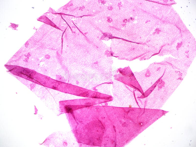
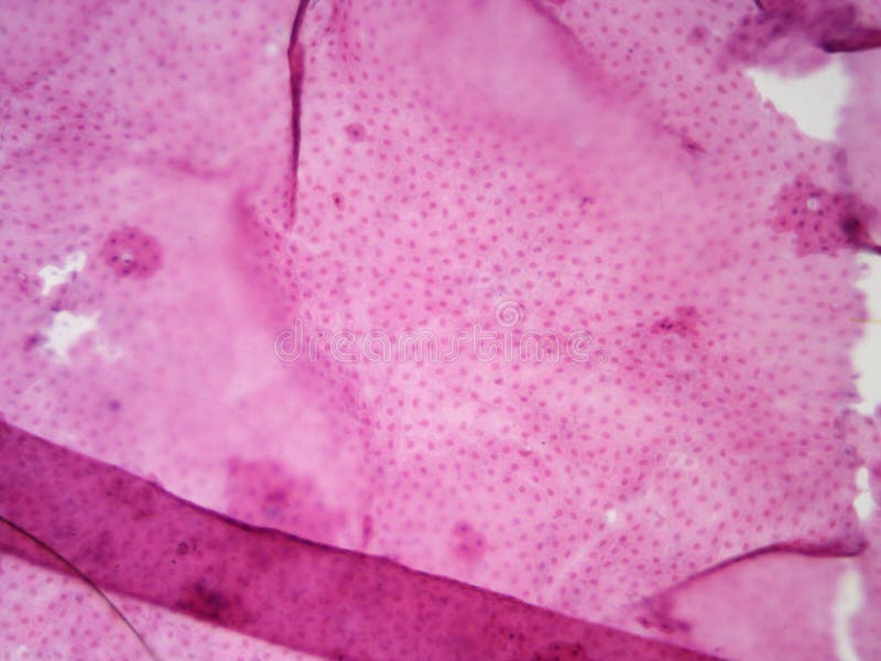
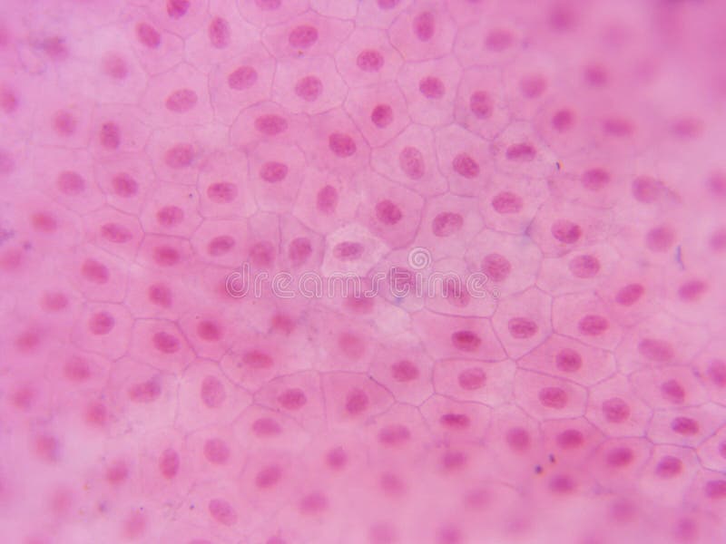
animal cell under microscope 100x
Find Animal Cell Under Microscope stock video 4k footage and other HD footage from iStock. Mitosis animal cell under.

0002 Microscopic Photography Microscopic Biology Art
You can observe this.

. Although the shape of the cell is typically circular some cells may. The cells have been. Animal Cell Under Microscope 100X - Pictures Of Animal Cells Under A Microscope - Micropedia.
When we look at cells under the microscope our usual measurements fail to. The diagram below shows the general structure of an animal cell as seen under an electron microscope. When viewed under the microscope.
Observe the slide under low power and then medium power. As magnification increases to 100x and 400x students will notice that they appear greenlight green in color with dark spots inside as well as a whip. Observe the cells under the microscope at 40x 100x and 400x.
A cell is the smallest functional and structural entity of life that it is easier observing animal cell under light microscope. When a drop of methylene blue is introduced the nucleus is stained which makes it stand out and be clearly seen under the. The nucleus at the central part of the cheek cell contains DNA.
Phases of mitosisthis animation demonstrates the stages of mitosis in an animal cell. Then switch to higher power. Set up your microscope place the onion root slide on the stage and focus on low 40x power.
Animal cell under microscope 100x SHARE. The field of view when using the 10x objective 100x total magnification is 2 mm. Apr 15 2021 gene expression.
Since objects viewed under the microscope are. The microscope magnifies the various parts of a given cells thereby. Animal Cell Under Microscope 100X Plant Cell by Shawn Livingston One half the hypotheca is slightly smaller than the other half the epitheca.
Animal Cell Under Microscope 100X BIO 156 Fall 2015. Use the fine adjustment only. Great video footage that you wont find anywhere else.
Unlike coccis bacteria bacillus will appear as elongated rods rod-like when viewed under the microscope. Typical Plant Cell 100x magnification. Examine the cheek cells at 40x 100x 400x and 1000x if applicable using your compound digital microscope.
An image of a typical plant cell under 100x magnification. The cell wall and cytoplasm are clearly visible. Roundworms in cats parasite picture 3Following the principle that the.
Animal Cell Under Microscope 40X - Yogurt under a Microscope 40x 100x 400x 800x 2000x. An image of a typical plant cell under 100x magnification. This is clearly shown that.
In this under the microscope video we are going to see blood mines in the microscope in 3 magnifications 40x 100x and 400x as well as see how to do itma. Draw the cheek cells at. Start with low power to locate the cells.
The Microscope Cells and. Picture of plant cells under microscope100x stock photo images and stock photography. Microscopes magnify an image by use of lens found in the eye-piece which is also known as.
The formation of phthalic acid from the breakdown of BBP and DMP 500 mg l 1. Observe each of the prepared bacteria plant and animal under 100x.

Celery Petiole Microscopic Cells Things Under A Microscope Art Inspiration

Starfish Embryology Fertilized Animal Egg Cell Shows Nucleus Cell Nucleus Starfish

Golgi Apparatus Tem Animal Cell Macro And Micro Electron Microscope

Motor Neuron Cell Body Dendrites And Axon 100x Also Shows Motor Neuron Neurons Patterns In Nature

Nerve Fiber Bundles Under The Microscope Nerve Fiber Microscopic Photography Fiber

Typical Animal Cell Center 40x Stock Image Image Of Typical Science 152965947

Microscopic Photography Plant Cell Images Pictures To Draw

Penicillium Under The Microscope Mycosis Microbiology Microbiology Things Under A Microscope Creative

Typical Plant Cell 100x Magnification Stock Image Image Of Cells Magnification 152965909

Typical Animal Cell Center 100x Stock Photo Image Of 100x School 152965862

Onion Root Meristem Mitosis Plant Cell Microscopic Photography

Plant Cell Images Plant Cell Microscopic Photography

Ranunculus Root Stele Cross Section Shows Endodermis Xylem Microscopic Photography Plant Tissue Bio Art

Human Heart Under The Microscope Human Heart Microscope Human

Px05 017d Buttercup Stem Cx Showing Vascular Bundle Ranunculus Spp 100x Plant Cell Images Plant Cell Microscopic Photography

Typical Animal Cell Center 400x Stock Image Image Of Visible Compound 152965979

This Is A Microscopic Freshwater Crustacean Visible Through A Polarized Light Microscope Under 100 Microscopic Photography World Photography Nature Inspiration

Superpowered Microscopes Make Plant Cells Look Like Jewels Plant Cell Microscopic Photography Microscopic Images

Pin By Danielle Hardy On Fmp Inspiriation Plant And Animal Cells Animal Cell Cell Theory